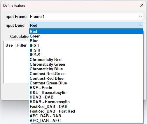
Bands

A band may be one of the standard red, green, blue channels, or it may be other image modalities. The different categories of bands are presented below.
RGB
The most used color model is the RGB-model (Red Green Blue). The RGB color space consists of the three additive primaries: red, green, and blue. Spectral components of these colors combine additively to produce a resultant color.
The RGB model is represented by a 3-dimensional cube with red green and blue at the corners on each axis. Black is at the origin. White is at the opposite end of the cube. The gray scale follows the line from black to white. In a 24-bit color graphics system with 8 bits per color channel, red is (255,0,0), green (0,255,0) and blue (0,0,255).
The RGB model simplifies the design of computer graphics systems but is not ideal for all applications. The red, green, and blue color components are highly correlated. This makes it difficult to execute some image processing algorithms. Many processing techniques, such as histogram equalization, work on the intensity component of an image only. These processes are easier implemented using the IHS color model.
In Visiopharm the band Red shows the red channel of the image, Green the green and Blue the blue.
IHS
The IHS (Intensity, Hue and Saturation) color model is a more intuitive color model to human vision, that separates color information from intensity information. The color is represented by the hue and saturation and the intensity determines the amount of light.
There are some variants of the IHS model. These includes HSB (Hue, Saturation, Brightness), HSV (Hue, Saturation, Value), HSL (Hue, Saturation, Lightness) and are not identical to the IHS model.
The IHS color space can be described as in the figure below. Here the hue is selected in the range 0-360° starting from the red primary located at 0°. The green primary is at 120° and the blue primary is at 240°. The pure colors (linear mixtures of adjacent pairs of red, green and blue) are located around the circumference with a saturation of 100%. Lowering the saturation value will result in a lighter color saturation. Intensity is in the axial direction, and increasing the value will result in a brighter color. The central vertical axis comprises the gray colors, ranging from black at intensity 0% to white at intensity 100%.
![IHS color model. [Source: Color image segmentation: advances and prospects, H.D.Cheng et al]](/img/classification/ihs_model.png)
Chromaticity
In the chromaticity space, a color is represented by the proportion of red, green and blue in the color, rather than by the intensity of each. Since these proportions must always add up to a total of 1, we are able to find just the red and green proportions of the color, and can calculate the blue value if necessary.
Although chromaticity contains less information than RGB color spaces, it has a number of useful properties for computer vision applications. As it is a measure of the relative intensity of each color component, chromaticity is more independent from variations in the lighting. Notably, where a scene viewed by a camera is not lit evenly - for example if lit by a spotlight - then an object of a given color will change in apparent color as it moves across the scene. Where color is being used to track an object in an RGB image, this can cause problems. The lack of intensity information in chromaticity images removes this problem, and the apparent color remains constant. Note that in the case where different parts of the image are lit by different colored light sources, problems can still emerge.
The band Chromaticity Red shows the red proportion of the image, Chromaticity Green the green proportion and Chromaticity Blue the blue proportion.
Can be very useful when trying to segment stainings with e.g. Toluidine Blue or Safranin O. In these cases one would use the Chromaticity Blue and Chromaticity Red respectively.
Contrast
These bands shows the difference between the two chosen color-bands. These bands are used if the contrast of two colors is more pronounced, than the intensity of a single color. The three contrasts found in Visiopharm are Contrast Red-Green, Contrast Red-Blue, and Contrast Green-Blue.
H&E
H&E stain is the most widely used stain in medical diagnosis. The terminology is based on the affinity to the dyes. The staining involves two staining procedure. First an application of hemalum, a complex formed from aluminium ions and oxidized hematoxylin, which colors the nuclei of cells blue. Second counterstaining with an aqueous or alcoholic solution of eosin, which colors eosinophilic structures red, these being cytoplasm, collagen etc. The H&E - Eosin band shows eosinophilic structures and H&E - Hematoxylin band shows the hematoxylin structures.
HDAB
HDAB is a custom defined input band which take hematoxylin and DAB (diaminobenzidine) staining into consideration by having the two stains (colors) as the primary and secondary axis in the color space coordinate system. Hematoxylin stain is an application of hemalum, a complex formed from aluminum ions and oxidized hematoxylin, which colors the nuclei of cells blue. DAB forms a brown color at the site of the target antigen or nuclei acid. DAB is a substrate chromogen system for use in peroxidase based immunohistochemical staining method. The HDAB - DAB band shows DAB structures and HDAB - hematoxylin band shows hematoxylin structures.
Fast Red DAB
FastRed_DAB is a custom defined input band which takes Liquid permanent red (LPR) and DAB (diaminobenzidine) staining into consideration by having the two stains (colors) as the primary and secondary axis in the color space coordinate system. LPR forms a red permanent reaction product at the site of the target antigen or nuclear acid. LPR is intended for use in immunohistochemical staining methods where alkaline phosphatase is the enzyme label. DAB forms a brown color at the site of the target antigen or nuclei acid. DAB is a substrate chromogen system for use in peroxidase based immunohistochemical staining method. The FastRed_DAB - DAB band shows DAB structures and the FastRed_DAB - FastRed band shows the LPR structures.
AEC DAB
AEC_DAB is a custom defined input band which take AEC (3-amino-9-ethylcarbazole) and DAB (diaminobenzidine) staining into consideration by having the two stains (colors) as the primary and secondary axis in the color space coordinate system. AEC forms a red color at the site of the target antigen or nuclear acid. AEC is a substrate chromogen system for use in peroxidase-based immunohistochemical staining method. DAB forms a brown color at the site of the target antigen or nuclei acid. DAB is a substrate chromogen system for use in peroxidase-based immunohistochemical staining method. The AEC-DAB - DAB band shows DAB structures and the AEC_DAB - AEC band shows the AEC structures.